BODY IMAGING CASE STUDIES
All of the internal disorders were found on thermograms that were taken for other reasons. Thermography was not used, and cannot be used, to screen for these conditions.
As with other healthcare examination procedures, all of the cases below were followed up with further tests in order to arrive at a final diagnosis.
The cases below demonstrate how thermography was able to help each patient find the cause of their health problem and ultimately get proper treatment.
CASE STUDY #1 – THYROID

The image of this patient’s neck shows a hyperthermic (hot) signal over the area of the thyroid. The patient presented with complaints that could be related to hypothyroidism. She had recently seen her doctor for her yearly physical and her labs were negative for a thyroid condition. She had been dealing with her symptoms for over 2 years, but her lab results were always normal. Considering the thermogram results, along with the patient’s symptoms, she was sent for an endocrinological consult. The endocrinologist ordered a significant complement of additional lab tests that confirmed that the patient was hypothyroid.
CASE STUDY #2 – DIABETES

The patient presented with neck pain. As per IACT guidelines, an upper body thermogram was done. The hand images showed infrared markers that are seen in some diabetics. A follow-up lab was done with the results positive for diabetes. In this case, the thermogram helped the patient get care for a condition that was going unnoticed.
CASE STUDY #3 -BACK PAIN

The patient presented with a history of low back pain radiating into the right leg. She had been undergoing physical therapy with only mild temporary relief. Her images showed infrared markers that suggested a possible disc compression of the nerve root at the L5/S1 level. The patient was sent for an MRI, which confirmed the diagnosis. The patient was finally able to get proper treatment for her condition.
CASE STUDY #4 – CARPAL TUNNEL SYNDROME

The patient presented with left thumb and shoulder pain due to falling on an outstretched hand. The infrared images of her hands showed thermal findings that suggested the possibility of median nerve pathology arising from the carpal tunnel. Follow-up tests confirmed that the patient had an early case of carpal tunnel syndrome. Rather than being treated incorrectly for an injury to the thumb, the patient was able to get proper care.
CASE STUDY #5 – DENTAL INFLAMMATION & STROKE WARNING

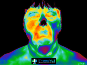
The patient in this case presented with neck and upper back pain. Her facial thermogram showed a display that suggested left sided dental inflammation and decreased blood flow in the terminal arteries of the left internal carotid artery. She was referred to her dentist who discovered an underlying infection. The patient was also sent for further imaging, which confirmed significant arteriosclerotic narrowing of the left internal carotid artery. In this case thermography acted as a risk marker for stroke. Since thermography was able to warn the patient of an increased risk of stroke, she was able to undergo surgery to remove the blockage in the artery.
CASE STUDY #6 – KNEE INJURY

The thermogram of this patient’s knees demonstrate an obvious hyperthermic (hot) left knee. The patient had sustained an injury to the knee while skiing. She had been treated for a sprain with some relief, but the pain had not resolved long after the injury. The patient had just received an orthopedic follow-up exam with no change in her diagnosis. Since the thermogram showed a significant amount of residual inflammation, the patient was sent for an MRI. The MRI showed that the patient had a complete rupture of the anterior cruciate ligament.
CASE STUDY #7 – SPINAL CONDITION

The patient in this case presented with chronic pain and tingling sensations isolated to the left lower leg. He had been treated conservatively for peripheral nerve entrapment with little to no changes in his symptoms. As per IACT guidelines, a lower body thermogram was performed. The thermogram showed a hypothermic (cold) distribution along the L4 and L5 thermatomes. This finding suggested that the problem existed at the spine. A follow-up MRI confirmed the spinal condition. The thermogram helped this patient to get the cause of his symptoms and the treatment he needed.
CASE STUDY #8
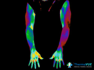
This thermogram shows an obvious hypothermia (cold) of the entire left arm. This patient had been experiencing chronic tingling and numbness. He had been diagnosed with a cervical spine neuropathy and had been undergoing physical therapy on and off with only mild temporary relief. The thermal findings were suggestive of a neurovascular problem likely arising from the thoracic outlet and not a cervical neuropathy. Upon further testing it was confirmed that the patient had thoracic outlet syndrome. With this new diagnosis the patient was able to get proper care.
CASE STUDY #9 – GETTING CORRECT DIAGNOSIS

This patient’s primary complaint was chronic right knee pain. She had seen her doctor and was referred for an orthopedic consult. A physical examination was performed along with x-rays. The orthopedist diagnosed her condition as osteoarthritis and recommended pain management as needed. Her thermogram shows a very focal area of hyperthermia (heat) directly over the medial aspect of her right knee. This area of inflammation is common with medial meniscus and/or medial collateral ligament damage. With this new information in-hand an MRI was ordered. The MRI confirmed the diagnosis as a torn medial meniscus.
CASE STUDY #10 – OSTEOARTHRITIS

This patient presented with pain in most of the joints in his hands. A physical examination suggested boney enlargement of both the proximal and distal interphalangeal joints (Bouchard’s and Heberden’s nodes). The thermogram demonstrates focal hyperthermic (hot) areas directly over these same joints (inflammation). X-rays were performed and confirmed the diagnosis as osteoarthritis.
CASE STUDY #11 – SYMPATHETIC NERVOUS SYSTEM & BLOOD CIRCULATION
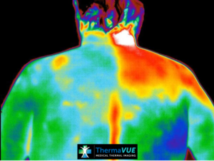
This patient presented with symptoms unrelated to what the thermogram is showing. For a separate health reason, the patient had previously undergone surgery to sever the sympathetic nerves leading out to the right neck, shoulder, and upper arm region. The thermogram demonstrates very clearly the sympathetic nervous system’s control over the blood circulation to the area. Without the sympathetic nervous system’s control over the circulation, the blood vessels remain fully open creating the hot red area seen on the thermogram. Notice the incredibly precise region of control extending from the nerves severed along the spine.
CASE STUDY #12 – RHEUMATOID ARTHRITIS

The patient’s primary complaint was pain in both knees and ankles. As an avid long-distance runner, the patient felt that his symptoms were simply a result of too much running. The thermogram showed perfect correlation with his symptoms – hyperthermia (hot) over both knees and ankles (inflammation). However, these thermal findings are not what are usually seen in injuries to long-distance runners. These thermal markers are more commonly seen in arthritis. As such, the patient was sent for lab testing. The lab results confirmed the diagnosis of rheumatoid arthritis.
CASE STUDY #13 – RESIDUAL INFLAMMATION – HEALING INCOMPLETE
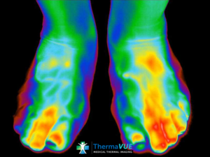
The patient initially complained of pain in the dorsal and plantar aspect of both feet and toes. As a collegiate level competitive runner, his presenting diagnosis was repetitive strain. He was placed on restricted training and underwent treatment with physical therapy. Before being released back to full training and competition, the patient was sent for a thermogram. The thermogram shown demonstrates a hyperthermic area (heat – inflammation) in both feet over the original areas of complaint. The image was invaluable in alerting the training and healthcare staff to the residual inflammation; and thus, incomplete healing of the patient’s injury. Without this information the athlete would have been placed back on the track prematurely with the possibility of causing severe injury.
CASE STUDY #14 – MYOFASCIAL PAIN AND DYSFUNCTION SYNDROME (MPDS)
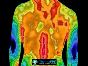
As a professional baseball player, this patient was being treated for a right shoulder impingement syndrome. After undergoing weeks of treatment with little to no improvement he was sent for a thermogram. The image shows focal areas of hyperthermia (heat) over the right rotator cuff. This thermal marker is commonly seen with myofascial pain and dysfunction syndrome (MPDS). A clinical exam correlated the findings and confirmed the diagnosis as MPDS. Within a week of treatment, the patient’s pain had significantly decreased along with a return of full range of motion in the shoulder. Thermography was instrumental in helping this patient find the cause of his condition.
CASE STUDY #15 – ACUTE GOUT
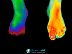
This patient came in with significant left foot pain. There was no history of trauma. The patient was an avid tennis player and wondered if this was the cause. The images showed infrared markers that were more common for gout. Follow-up lab tests confirmed that the patient had gout.
CASE STUDY #16 – SINUS INFLAMMATION
