BREAST IMAGING CASE STUDIES
NB. Just as with many other healthcare examination procedures, all of the cases below were followed up with further tests in order to arrive at a final diagnosis.
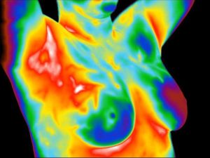
The cases below clearly demonstrates the major role thermography plays as a breast cancer risk assessment marker.
BREAST CASE STUDY #1
This patient presented with a lump in the right breast.
Her doctor sent her in for a follow-up mammogram to investigate the lump.
The mammogram came back normal with a recommendation for a routine annual exam.
IMAGE A

A hot vascular pattern (angiogenesis) is shown projecting into the area of the lump in the upper outer quadrant of the right breast.
IMAGE B

The left breast is shown to be cool with a normal limited vascular pattern.
IMAGE C

The right breast showing the area of the lump directly facing the infrared detector. The increased heat and vascularity are very noticeable especially when compared to the left breast.
The patient was sent back to her doctor with a recommendation for further testing.
A biopsy confirmed that the lump was cancer.
BREAST CASE STUDY #2
This patient had a recent mammogram that was considered watchful for an area in the left breast.
A follow-up ultrasound of the left breast was also watchful for the same area.
The report recommended a follow-up mammogram in 6 months to monitor the finding in the left breast.
IMAGE A

A noticeable increase in heat and blood vessel activity (angiogenesis) is seen in the left breast when compared to the right.
IMAGE B

The right breast shows a normal cool thermal signal and vascular pattern.
IMAGE C

The left breast is viewed showing the extent of the hot engorged blood vessels extending across the breast.
The patient was sent back to her doctor with a recommendation for further testing with a focus on the watchful area in the left breast.
A biopsy came back positive for cancer.
BREAST CASE STUDY #3
Estrogen Dominance.
This patient presented with pain and tenderness in both breasts.
IMAGE A

The image above shows a display of large hot blood vessels extending across the upper half of both breasts (You may also notice that there is more going on in the left breast, but for now we will focus on these other findings.).
This thermovascular display is commonly seen in patients with a suspected estrogen dominant state of the breasts.
Since the greatest single risk factor for the development of breast cancer is lifetime exposure to estrogen, this finding may be very important in the prevention of breast cancer.
In order to confirm the condition, the patient was referred to her doctor for an examination and further testing. Her follow-up exam and testing confirmed the estrogen dominance.
IMAGE B

After undergoing treatment, the image above shows a significant decrease in blood vessels, blood vessel size, and heat across the breasts.
You may also notice the overall improvement in the left breast.
The patient’s left breast was a TH4 (abnormal) in the first image (Image A) and is now a TH3 (questionable).
The patient underwent specific treatment for her estrogen dominance and separate treatment for her TH4 breast.
Her images show significant improvement with near normalization of the hormone balance in her breasts.
Her previous symptoms of pain and tenderness had also resolved.
Thermography was able to warn this patient of her increased risk for breast cancer due to the estrogen dominance.
With this information in-hand she was able to get the treatment she needed to lower her risk for future breast cancer.
BREAST CASE STUDY #4
This case demonstrates how angiogenesis is involved in malignant tumor growth and how infrared imaging detects this process.
IMAGE A

The image above shows a significant amount of heat and blood vessels in the left breast.
The right breast is cool and normal in comparison.
The patient was sent back to her doctor for immediate further imaging.
The combined findings on the thermogram and structural imaging prompted her doctor to order a biopsy.
The biopsy was positive for a very early stage cancer.
IMAGE B

This image was taken three months after the patient’s lumpectomy.
Note how the left breast has returned to a near normal cool state.
The blood vessels seen in the previous image have almost vanished.
Since the cancer has been removed, there is no longer a demand for an increased blood supply to maintain the continued growth of the tumor.
BREAST CASE STUDY #5
This patient had normal mammogram and a watchful ultrasound of the left breast.
The report recommended follow-up imaging in 6 months.
Her doctor was concerned over a thickening in the left breast.
IMAGE A

A significant increase in temperature of the entire left breast along with noticeable vascularity (angiogenesis) is shown.
IMAGE B

The right breast is seen to be normal and cool without evidence of abnormal vascular patterning.
IMAGE C

The image shows the extent of thermovascular activity across the left breast.
The patient was referred to her doctor with the recommendation for further testing.
The biopsy results were positive for cancer.
BREAST CASE STUDY #6
This case study involves a 22-year-old woman who discovered a lump in her right breast.
Her doctor examined the area and noted that it was likely a cyst.
The patient was concerned and asked for further testing.
A mammogram was performed with normal findings.
Her doctor assured her that everything was fine and that she was too young to have breast cancer.
IMAGE A

The image shows an increase in temperature and vascularity (angiogenesis) in the right breast.
IMAGE B

The image shows that the left breast is cool and without a suspicious vascular pattern.
IMAGE C

This image of the right breast shows a group of hot blood vessels over the area of the lump.
The patient was referred to her doctor for further testing.
A biopsy confirmed that the lump was a very early stage cancer.
BREAST CASE STUDY #7
This 41-year-old woman presented with a complaint of pain in the left breast.
Her last physical examination and mammogram one year earlier were normal.
IMAGE A

The image shows a increase in temperature and vascularity (angiogenesis) in the left breast.
IMAGE B

This image shows the right breast is relatively cool and with no suspicious vascular patterns.
IMAGE C

